Latest Features
Spinal DCS: A Personal and Professional View
When a diving physician becomes his own patient. Andrew Pitkin recounts his brush with spinal decompression sickness — blending firsthand fear, clinical insight, and the hard truths that data alone can’t explain.
By Dr. Andrew Pitkin
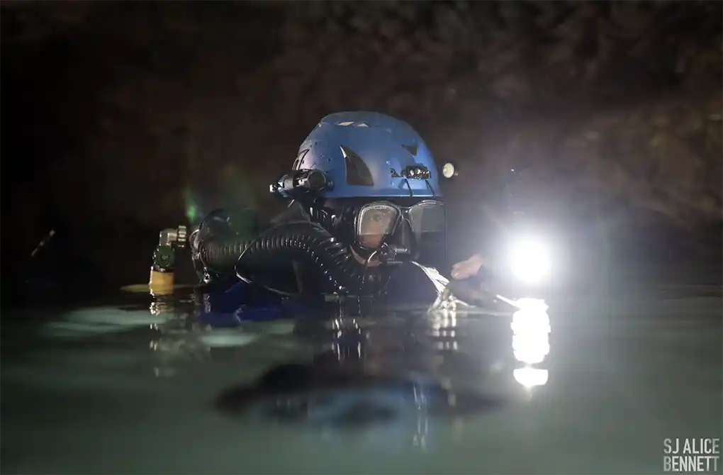
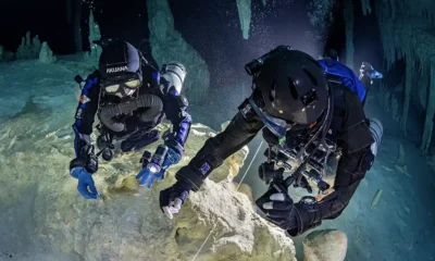
Lead Image: The author preparing to dive. Photo by SJ Alice Bennett.
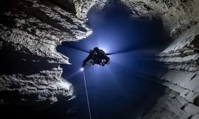
In October 2023, I experienced the first-ever episode of decompression sickness (DCS) of my thirty-year technical diving career. During the decompression from my deepest dive—to 174 m/570 ft—I started experiencing back pain at the 51 m/170 ft decompression stop. I was used to experiencing lumbar discomfort during long dives, especially when using a diver propulsion vehicle (DPV) over long distances; but this pain was higher up in the lower thoracic region. Initially confined to the midline and just to the right of it, the pain gradually got worse and spread to the left side. It didn’t seem to be affected by movement or breathing.
Being a trained diving physician, I became suspicious that this odd discomfort might be the harbinger of spinal cord DCS. So I began wiggling my toes and trying to decide if I had any neurological impairment in my legs—no easy feat in a drysuit. I also thought about what to do if this was what I feared. I knew that, if I had developed spinal DCS symptoms at this depth, continued to ascend, and completed another 10 hours of planned decompression, the likeliest outcome would be a severe spinal injury that could easily leave me paraplegic and incontinent, even with prompt recompression.
At this remote dive site, we were at least four hours from the nearest chamber (by helicopter), but I also had to dive through over a mile of shallow (less than 15 m/50 ft) undulating underwater cave passage to get back to the entrance for evacuation. I might have to do it without the use of my legs. I also didn’t have a buddy to help me, but that’s another story.

These grim circumstances churned through my mind over the next few minutes. As I was still optimistically searching for an alternative explanation for the back/lower chest pain, a wave of painful paraesthesiae (“pins and needles”) washed over the entire lower half of my body, down both of my legs, and was most severe in the soles of my feet. I couldn’t immediately detect any weakness, but I wasn’t waiting to find out.
I grabbed my DPV and started scootering deeper into the cave. The passage here descends at a 45-degree angle to a depth of 72 m/240 ft, then ascends about 12 m/40 ft, before continuing its descent to 100 m/330 ft where it levels off. I landed on the rocks at 72 m/240 ft and laid there, focused on the pain in my back and the sensations in my legs. I kept the DPV handle in my hand, ready to go down to 100 m/330 ft if the symptoms didn’t start to improve quickly.
Being a victim of this disease created a desire to do a deep knowledge dive into the subject of spinal DCS. Below, I’ll discuss spinal DCS in some detail, review what is known about it scientifically, and explore why this particular form of DCS stands out as a particularly strange and malignant variant.

The clinical picture
Since decompression sickness was first encountered, its ability to cause a devastating injury to the spinal cord has been obvious. The classic picture, familiar to all diving physicians, is the early onset of lower thoracic back pain which often spreads circumferentially around the trunk (so-called “girdle pain”) and is typically associated with sensory symptoms and weakness in the legs. Balance and walking are often profoundly affected due to motor weakness and involvement of the nerves responsible for proprioception (i.e., how the brain knows where the parts of the body are). In more severe cases, the diver has difficulty with bladder or bowel control.
The clinical course is highly variable; many cases improve transiently with normobaric oxygen before deteriorating with sometimes much more severe manifestations than the initial symptoms. Such cases are often the ones left with the most severe residual neurological deficits.
It has also been known for many decades that, even with relatively prompt (within a few hours) recompression treatment, spinal DCS can leave the diver with a permanent neurological deficit. In severe cases, this may affect their ability to walk unaided and maintain bladder and bowel control—devastating and potentially life-shortening outcomes. A number of studies have confirmed the clinical impression that the diver’s residual neurological deficit is more closely related to the severity of the disease than any differences in treatment.

In 1996, Boussuges and colleagues published a scoring system for severity of neurological decompression sickness [1] in which the clinical features most associated with an adverse outcome were those of spinal DCS (paraplegia and urinary disturbances), along with deteriorating rather than improving or unchanging symptoms before recompression (see the table below). A few years later, I and my colleagues at the Institute of Naval Medicine in the UK published an analysis of British diving accidents in which we applied Boussuges’s scoring system; we found that cases with a score greater than 7 were likely to have a long-term functionally-important neurological deficit, whereas those that scored 7 or less were very unlikely to [2].
In 2010, Jean-Eric Blatteau and colleagues retrospectively analyzed data from 279 cases reported from seven hyperbaric centers in France and Belgium and found that, like in the British cohort, a Boussuges severity score greater than 7 was associated with a poor outcome. Importantly, they found that neither time from symptom onset to recompression nor the choice of hyperbaric treatment table had a significant influence on the final outcome [3].
Another study by the same group subsequently found the same result in military divers who had been recompressed within six hours of symptom onset [4]. A large study of 5,278 cases of DCS treated with recompression in China found a similar correlation between severity and outcome, although they found that delay to recompression did also have a small effect [5].

Put simply, the evidence is that the primary determinant of recovery after spinal DCS is how severe the episode is, not how quickly it is treated. One caveat is that these retrospective studies may be biased in that more severe cases are likely to be treated earlier, thereby tending to reduce any effect of time to treatment on outcome.

Magnetic resonance imaging in spinal DCS
Magnetic resonance imaging (MRI) is by far the most useful method of imaging the spinal cord. Gempp and colleagues analyzed MRI images of 45 divers with spinal DCS to whom they also applied Boussuges’s severity score [6]. 48% of the divers had a score greater than 7 and 86% of these had incomplete resolution of symptoms, compared with only 8% of the divers with a score of 7 or less. 31% of divers had abnormalities (hyperintensities on T2-weighted images) in the posterior or lateral white matter of the cord, and these were much more common in divers with a severity score above 7.
The abnormalities typically correlated with the clinical picture and occurred mostly in the cervical and thoracic parts of the spinal cord. Interestingly, one-third of the divers also had degenerative disease of the vertebrae or intervertebral disks and, in these cases, the site of contact with the cord correlated with at least one of the observed hyperintensities in the cord. There was no statistical correlation between vertebral disease and outcome, but this observation raises some very interesting questions.
A more recent study published by Hennidge and colleagues reviewed MRI images from seven patients with spinal DCS [7]. Six of these had diverse T2-weighted abnormalities in the thoracic and cervical cord, and five of these also had involvement of the gray matter, a finding which the authors (possibly incorrectly) attributed to arterial bubble emboli.

The pathology of spinal DCS
It is now universally accepted that DCS is caused by bubble formation. Although other mechanisms have been proposed, it is the transition of dissolved inert gas into the gaseous phase in the body that has harmful effects, and these may result in clinical symptoms. DCS has many different forms; in many cases, how bubbles cause the symptoms they do is, unfortunately, still poorly understood. For an overview, I would suggest Simon Mitchell’s excellent review article published in 2024—in particular, his Table 1, which summarizes what is known or theorized about how bubbles cause symptoms in different organ systems [8].
In the case of the spinal cord, the mechanism of DCS has been debated for many decades. In 1908, Boycott, Damant, and Haldane first proposed arterial bubbles as a mechanism in their classic paper on decompression sickness. In the same paper, they also conjectured that, because of the high lipid content and poor blood supply of the spinal cord white matter, any bubbles that lodge there would enlarge from gas diffusion from nearby tissue [9].
However, it has always seemed unlikely that arterial bubbles were the primary cause of spinal injury in diving because the brain receives a much larger blood supply (about 80 times more) than the spinal cord; yet, it is rarely affected in cases of severe spinal DCS, although some studies have shown a correlation between spinal DCS and a right-to-left intracardiac shunt, such as that through a PFO [10].

This is a very different picture compared to other diseases of embolic phenomena (such as arterial gas embolism from pulmonary barotrauma and paradoxical venous thromboembolism or fat embolism) where the brain is the primary target, and the spinal cord is almost never affected. A consistent finding from animal and autopsy studies of spinal DCS is that the white matter of the cord (containing nerve fibers) is far more affected than the gray matter—where the nerve cell bodies are, and which receives the majority of the blood supply.
A second, and possibly most popular, theory is that the spinal cord is susceptible to having bubbles form in situ (so-called autochthonous bubbles) because the myelin (a physiological electrical insulator) has a high lipid (fat) content and therefore dissolves inert gas readily [11]. Research published by Francis and colleagues using an experimental model of spinal DCS in dogs showed numerous space-occupying lesions (presumably caused by bubbles) mostly in the white matter. However, the spaces, which measured between 20 and 200 µm in diameter, did not seem to be large enough on their own to account for the amount of measured neurological deficit [12].
This does not argue against the mechanism, since secondary injury from inflammation, hemorrhage, and edema is highly likely; Broome and colleagues demonstrated multifocal hemorrhagic lesions (see picture) in a porcine model of severe spinal DCS which correlated with functional outcome [13].

The autochthonous bubble mechanism has some limitations. The animal models supporting it used extremely provocative dive profiles to cause pathology that could be studied, and in situ bubble formation could not be generated without a nitrogen supersaturation of 3.6 ATA [14], a degree of supersaturation rarely encountered during normal diving. The white matter of the cord, with limited perfusion and a high inert gas solubility, would be expected to absorb and release inert gas relatively slowly (i.e., it is a “slow” tissue) [15], which is inconsistent with the typical short latency of spinal DCS. Spinal DCS is almost never seen in saturation decompression or altitude exposure, and its strong association with short hyperbaric exposures and rapid ascents (such as in indigenous fishermen) suggests that “fast” tissue compartments are responsible.
It has also been suggested that so-called autochthonous bubbles are actually trapped venous bubbles [16]. This leads neatly into a third mechanism proposed in 1955 by Haymaker and Johnston who suggested that obstruction to venous drainage of the spinal cord by bubbles could be the mechanism of injury [17]. The venous system of the cord is anatomically complex and variable between individuals. It consists of a longitudinal network of valveless veins first described by French anatomist and physician Gilbert Breschet in 1819 and now commonly known as Batson’s plexus. Oscar V. Batson was an American otolaryngologist who, in 1940, showed that the spinal cord’s venous drainage was a low-pressure, large-capacitance network without valves that could be responsible for the spread of certain cancers (e.g. prostate) to the spine [18].
The proposed mechanism for its role in decompression sickness is that a rise in central venous pressure caused by immersion (and sometimes augmented by venous bubble emboli from other tissues arriving in the pulmonary arteries) could cause engorgement of the plexus, making it a likely site of bubble accumulation because of the large capacity of the network and the slow movement of inert gas-rich blood within it. Both the bubbles themselves and secondary effects such as clots could thereby obstruct venous drainage of the spinal cord. This is known to cause damage (so-called venous infarction). However, because of the extensive connections of Batson’s plexus to other parts of the venous system, it is rarely seen in other contexts; but when it does, the clinical picture is very similar to spinal DCS [19].

Bottom arrow – spinal cord
This mechanism has a considerable amount of evidence to support it. In a well-designed series of experiments, Hallenbeck, Bove, and Elliott demonstrated clear evidence of epidural venous obstruction in an animal model of spinal DCS [20]. Shortly after venous bubbles were detected in the heart by Doppler ultrasound, there was a large rise in cerebrospinal fluid (CSF) pressure (see table) with only a small rise in central venous pressure, a finding that strongly suggests severe obstruction to venous drainage of the spinal cord, since CSF drains into these veins via the arachnoid villi. They were able to show—with limited 1970s imaging techniques—that the epidural venous plexus was obstructed, distended, and contained moving and static filling defects (almost certainly bubbles). At autopsy, extensive white matter hemorrhage was seen—most commonly in the thoracic region, less often in the cervical region—and extensive thrombosis (clots) were found in the epidural venous system. These findings are exactly the same as those in humans with severe spinal DCS [21].
| CSF pressure (mm H2O) | Pre-dive control | Post spinal cord involvement |
| Mean | 80 | 524* |
| Range | 41 – 163 | 218 – 816 |
Cerebrospinal fluid (CSF) pressure changes during decompression sickness in 8 dogs studied at Duke University. *Difference is significant at p < 0.0025 by paired t-test.
The venous infarction hypothesis is attractive: it could explain all the known features of spinal DCS, including its patchy diffuse localization, relapsing and remitting course, resistance to recompression treatment, but especially the odd vulnerability of the spinal cord compared with the brain, which has a different route of venous drainage.
It is possible that the high level of perfusion of the brain (making it a “fast” tissue) allows it to contribute venous gas (rich in inert gas or even bubbles) to Batson’s plexus via connections at the base of the skull, thereby explaining the short latency of spinal cord involvement. The venous obstruction will impair removal of inert gas and promote or even initiate autochthonous (in situ) bubble formation as a secondary phenomenon.
In addition, the acute rise in CSF pressure (and, therefore, intracranial pressure) could very neatly account for diffuse CNS manifestations such as headache, nausea, visual disturbances, lethargy, confusion, and unconsciousness—all features that have been observed in fulminating spinal decompression sickness. It is also easy to imagine how mechanical compression of Batson’s plexus by degenerative spine disease could promote local bubble and clot formation, explaining the correlation seen in one MRI study.
Another piece of supporting evidence comes from thoracic surgery. Repair of a diseased aorta in the chest can compromise the arterial blood supply of the spinal cord, and a commonly-employed technique to reduce the risk of a spinal injury is to artificially lower CSF pressure [22]. It is well-known that an elevated spinal venous (and therefore CSF) pressure dramatically increases the vulnerability of the cord to compromised arterial perfusion.
By analogy, any injurious effects of arterial bubbles are likely to be considerably amplified if the CSF pressure is elevated, as shown in Hallenbeck and colleagues’ studies. While the other two mechanisms of arterial bubble embolization and autochthonous bubble formation may still occur, they can clearly be amplified by epidural venous obstruction or even be caused by it, and neither can explain all the known features as completely.

What doesn’t work, doesn’t work
Like many technical divers, I use gradient factors to add conservatism to the parallel compartment decompression model programmed into my dive computers. Because of emerging scientific evidence over the past two decades, I had been gradually changing the gradient factors I used in order to shift some of my total decompression time from deeper decompression stops to shallower ones [23]. The 2011 publication of the results of the US Navy’s study comparing a deep stops profile with one using shallow stops for the same total decompression time [24] was largely responsible, although other supportive evidence was already accumulating. For anyone interested, I was using gradient factors of 65/85 on my dive.
When symptoms occur during in-water decompression during a dive (that has otherwise gone as planned) the reason is clear: decompression up to that point has been inadequate and should have included deeper stops or a slower ascent rate. My incident prompted a re-evaluation of the evidence surrounding deep stops, including that provided by the NEDU study.
“I suggest there is a flaw, not in the evidence itself, but in how we as technical divers may have used it to inform our diving. As I see it, the main problem is the equal weighting of all DCS cases.”
This is an appropriate choice for a decompression researcher and, as a diver, I naturally want to minimize my chance of any DCS. But not all cases of DCS are the same; some skin itching or limb pain, both certain to disappear over time with no treatment, are almost irrelevant compared with the potentially severe consequences of spinal DCS: paraplegia, loss of control of bladder and bowel function, and inability to walk normally (or at all) for the rest of one’s life. If spinal DCS seems likely to be prevented by a deep stop strategy, and milder type 1 symptoms may be increased by it (there is evidence to support this), the decision on the use of deep stops should be weighted according to the consequences of each. In my mind, that weighting should be very heavy indeed during a high-risk deep dive: why should I care if I get some arm pain or itching if it helps me avoid being paralyzed? And, as discussed above, even prompt recompression in a chamber may well not achieve full resolution in severe cases.
Of course, there is no guarantee that a deep stops strategy will prevent spinal DCS; indeed, the only case (to my mind) of DCS in the NEDU study that looked like a spinal episode was in the deep stops group (see Appendix D, dive day 28). I believe this is a typical spinal DCS case, with features that mesh very nicely with the pathological mechanisms discussed earlier. The diver’s symptoms started within 10 to 15 minutes of surfacing and worsened rapidly. His “dull, bilateral posterior trapezius muscle pain” was clearly typical girdle pain and his patchy and progressive neurological deficit with gait disturbance and ataxia were characteristic of spinal involvement. The nausea, vomiting, visual disturbances, and headache are difficult to explain with a pure spinal cord injury but are beautifully consistent with the early and substantial rise in intracranial pressure found by Hallenbeck and colleagues in their experiments in the early 1970s. Clearly, that decompression schedule on that day was not sufficient to prevent spinal DCS in that diver, and it is impossible to know whether the incident would have been more or less severe if he had used the shallow stop schedule.

The outcome
Back to my dive.
To my immense relief, both the girdle pain and the paraesthesiae started diminishing almost immediately once I reached a depth of 72 m/240 ft. Over the next two minutes, the sensory symptoms gradually disappeared, and I was left only with a slight residual ache where the back pain had first appeared. I decided to wait at that depth for 15 minutes before slowly ascending 3 m/10 ft while monitoring myself very carefully for any recurrence of the symptoms. I spent about 10 minutes at that next stop, and then repeated this every 3 m/10 ft up to the 48 m/160 ft depth where the symptoms had originally appeared. I was obviously concerned that the symptoms might recur there, but I was able to continue decompressing, essentially by guesswork, until I caught up with my dive computer schedule at 21 m/70 ft. The computer had added a lot of shallow decompression time because of my extended stay at depth; I was able to spend 6 hours of it in comfort in a habitat before making my way back to the entrance where I surfaced after 16.5 hours underwater—about 4 hours longer than if nothing had happened (see dive profile below).

After the dive, I had no residual symptoms that I could detect. But, for the next few mornings when I woke up, I was aware that the sensation of the bedsheet touching the soles of my feet was not quite normal. Neurologically, this modality is known as ‘light touch,’ and the abnormality disappeared (or I couldn’t detect it) once I got up and started walking around. After three or four days, this last vestige of the incident had gone.
There was no doubt in my mind that I had been incredibly lucky. Over the following year, I heard of several incidents of spinal DCS affecting people I knew, and they had a number of features in common: very deep dives, older divers like me (I am in my fifties), and symptoms appearing during decompression. In diving medicine, the interval between surfacing and symptom onset is termed latency and, in general, the shorter the latency the more severe the DCS. These episodes all had negative latency: onset during decompression. In the other cases I heard of, all the divers re-descended at least once with varying degrees of improvement, and most still had symptoms present or recurring after surfacing and were treated in a hyperbaric chamber. It is possible that this cluster of spinal DCS is occurring by chance—in a population of technical divers who are going deeper and who are becoming more susceptible as they get older.
“Decompression procedures for very deep dives cannot necessarily be extrapolated from what works during shallower ones.”
I was fortunate that I have no residual neurological deficits. Some of my deep diving friends and acquaintances have not been so lucky. Comparing our dive profiles, it appears that my re-descent at the onset of symptoms was relatively deeper and more prolonged, and whether this is the reason that I escaped more serious injury is unknowable. It is clear that to continue to ascend with significant spinal symptoms is probably going to be disastrous, however unappealing the alternative is. Immediate descent as soon as symptoms occur might be the best way of dealing with this malignant form of spinal DCS where symptoms start during the decompression and, if so, the logistic consequences of doing so should be incorporated into dive planning. Once on the surface, the diver should immediately seek emergency hyperbaric medical advice because of the high likelihood of relapse.
Extreme dives such as this are so far outside of the testing envelope of decompression models that they must be considered experimental. A few anecdotal cases are not good grounds for altering decompression procedures for shallower dives, but they provide some evidence of the failure of those decompression models when extrapolated to much greater depths. Symptoms arising during decompression—particularly at substantial depth as in my case—suggest that a slower ascent to that point, in the form of more deep stops, is required. This could be achieved by decreasing the low gradient factor or by some other empirical method, such as ‘Pyle’ stops [25]. While anecdotal evidence is the weakest kind, all new science begins with observations, especially those that do not fit in with established theory.

Spinal cord decompression sickness is one of the most severe and potentially life-threatening forms of this bizarre disease, and much research has been carried out into its pathology. The venous infarction hypothesis appeals to me the most because of its ability to explain every feature more completely than the traditional in situ bubble formation and arterial embolization mechanisms. Whether this model of pathogenesis leads to better prevention and treatment remains to be seen.
“All divers—especially middle-aged ones undertaking extremely deep dives—would benefit from considering the risks and benefits of a deep stop strategy and weighing them according to the severity of the consequences: paralysis or limb pain? The choice may be yours.”

References
- Boussuges A, Thirion X, Blanc P, Molenat F, Sainty JM. Neurologic decompression illness: a gravity score. Undersea Hyperb Med. 1996 Sep;23(3):151-5. PMID: 8931282.
- Pitkin AD, Benton PJ, Broome JR. Outcome after treatment of neurological decompression illness is predicted by a published clinical scoring system. Aviat Space Environ Med. 1999 May;70(5):517-21. PMID: 10332950.
- Blatteau JE, Gempp E, Simon O, Coulange M, Delafosse B, Souday V, Cochard G, Arvieux J, Henckes A, Lafere P, Germonpre P, Lapoussiere JM, Hugon M, Constantin P, Barthelemy A. Prognostic factors of spinal cord decompression sickness in recreational diving: retrospective and multicentric analysis of 279 cases. Neurocrit Care. 2011 Aug;15(1):120-7. doi: 10.1007/s12028-010-9370-1. PMID: 20734244.
- Blatteau JE, Gempp E, Constantin P, Louge P. Risk factors and clinical outcome in military divers with neurological decompression sickness: influence of time to recompression. Diving Hyperb Med. 2011 Sep;41(3):129-34. PMID: 21948497.
- Xu W, Liu W, Huang G, Zou Z, Cai Z, Xu W. Decompression illness: clinical aspects of 5278 consecutive cases treated in a single hyperbaric unit. PLoS One. 2012;7(11):e50079. doi: 10.1371/journal.pone.0050079. Epub 2012 Nov 21. PMID: 23185538; PMCID: PMC3503765.
- Gempp E, Blatteau JE, Stephant E, Pontier JM, Constantin P, Pény C. MRI findings and clinical outcome in 45 divers with spinal cord decompression sickness. Aviat Space Environ Med. 2008 Dec;79(12):1112-6. doi: 10.3357/asem.2376.2008. PMID: 19070307.
- Hennedige T, Chow W, Ng YY, Chung-Tsing GC, Lim TC, Kei PL. MRI in spinal cord decompression sickness. J Med Imaging Radiat Oncol. 2012 Jun;56(3):282-8. doi: 10.1111/j.1754-9485.2012.02386.x. Epub 2012 May 25. PMID: 22697324.
- Mitchell SJ. Decompression illness: a comprehensive overview. Diving Hyperb Med. 2024 Mar 31;54(1Suppl):1-53. doi: 10.28920/dhm54.1.suppl.1-53. PMID: 38537300; PMCID: PMC11168797.
- Boycott AE, Damant GC, Haldane JS. The Prevention of Compressed-air Illness. J Hyg (Lond). 1908 Jun;8(3):342-443. doi: 10.1017/s0022172400003399. PMID: 20474365; PMCID: PMC2167126.
- Gempp E, Blatteau JE, Stephant E, Louge P. Relation between right-to-left shunts and spinal cord decompression sickness in divers. Int J Sports Med. 2009 Feb;30(2):150-3. doi: 10.1055/s-2008-1038844. Epub 2008 Sep 4. Erratum in: Int J Sports Med. 2009 Feb;30(2):153. PMID: 18773377.
- Bond JP, Kirschner DA. Spinal cord myelin is vulnerable to decompression. Mol Chem Neuropathol. 1997 Apr;30(3):273-88. doi: 10.1007/BF02815103. PMID: 9165491.
- Francis TJ, Griffin JL, Homer LD, Pezeshkpour GH, Dutka AJ, Flynn ET. Bubble-induced dysfunction in acute spinal cord decompression sickness. J Appl Physiol (1985). 1990 Apr;68(4):1368-75. doi: 10.1152/jappl.1990.68.4.1368. PMID: 2347778.
- Broome JR, Dick Jr EJ, Dutka AJ. Multifocal CNS haemorrhage as a cause of recompression treatment failure in a pig model of neurological decompression illness. Undersea Biomed Res. 1994;21:69-70.
- Francis TJR. The role of autochthonous bubbles in acute spinal cord decompression sickness (PhD thesis). London: University of London; 1990.
- Hempleman HV. History of Evolution of Decompression Procedures. In: Bennett PB, Elliott DH eds. The Physiology and Medicine of Diving, 3rd ed. London: Bailliere Tindall; 1982.
- Palmer AC. Nature and incidence of bubbles in the spinal cord of decompressed goats. Undersea Hyperb Med. 1997 Sep;24(3):193-200. PMID: 9308143.
- Haymaker W, Johnston AD. Pathology of Decompression Sickness: A Comparison of the Lesions in Airmen with those in Caisson Workers and Divers. Military Medicine 1955 Sept; 117(3): 285–306.
- Batson OV. The vertebral vein system, Calldwell lecture, 1956. Bone Metastasis. 1981:21-48.
- Kim RC, Smith HR, Henbest ML, Choi BH. Nonhemorrhagic venous infarction of the spinal cord. Ann Neurol. 1984 Apr;15(4):379-85. doi: 10.1002/ana.410150413. PMID: 6742784.
- Hallenbeck JM, Bove AA, Elliott DH. Mechanisms underlying spinal cord damage in decompression sickness. Neurology. 1975 Apr;25(4):308-16. doi: 10.1212/wnl.25.4.308. PMID: 1168317.
- Kitano, M., Hayashi, K., & Kawashima, M. Three autopsy cases of acute decompression sickness. J West Jap. Orthop. Traum. 1977; 26(3) : 110-116.
- Coselli JS, LeMaire SA, Köksoy C, Schmittling ZC, Curling PE. Cerebrospinal fluid drainage reduces paraplegia after thoracoabdominal aortic aneurysm repair: results of a randomized clinical trial. J Vasc Surg. 2002 Apr;35(4):631-9. doi: 10.1067/mva.2002.122024. PMID: 11932655.
- Doolette DJ. Gradient factors in a post-deep stops world. inDepth May 29, 2019.
- Doolette DJ, Gerth WA, Gault KA. Redistribution of decompression stop time from shallow to deep stops increases incidence of decompression sickness in air decompression dives. Technical Report. Panama City (FL): Navy Experimental Diving Unit; 2011 Jul. 53 p. Report No.: NEDU TR 11-06.
- Pyle RL. The importance of deep safety stops: Rethinking ascent patterns from decompression dives. DeepTech 1996; 5:64


Enjoyed this read? Here are more stories that tie into it.
DIVE DEEPER
InDEPTH: Decompression Habitats Are Ascendent By Andy Pitkin (2022)
InDEPTH: When Easy Doesn’t Do It: Dual Rebreathers in Extended-Range Cave Diving by Tim Blömeke (2022)
Diving Hyperb Med: Is more complex safer in the case of bail-out rebreathers for extended range cave diving? Derek B Covington, Charlotte Sadler, Anthony Bielawski, Gareth Lock, Andrew Pitkin (2016)
InDEPTH: Rebreather Forum 4 Proceedings (2023)

Andrew Pitkin learned to dive in 1992 in the cold, murky waters of the United Kingdom; he started cave and technical diving in 1994. From 1996 to 2000, he was employed as a civilian diving physician at the Royal Navy’s Institute of Naval Medicine, running a hyperbaric facility, treating decompression illness, and participating in research related to post-decompression illness outcomes, submarine escape, and military underwater breathing system testing. He moved to the USA in 2007 and is currently on the faculty of the College of Medicine at the University of Florida in Gainesville, Florida, where he has participated in numerous underwater cave exploration and filming projects. His background in hyperbaric medicine and anesthesiology, combined with his interest in electronic and mechanical engineering, has resulted in the development of innovative equipment for technical cave exploration, including various rebreathers, suit heater controllers, improved DPV battery packs, heads-up displays for rebreathers, single-wire telephones, and underwater radiolocation systems.
Links:
Website: https://www.karstunderwater.org
YouTube: https://www.youtube.com/@andrewpitkin5243
UF Health Bio: https://ufhealth.org/doctors/andrew-d-pitkin/bio


From Fear to Fieldwork: How Science Rewrites the Shark Story

By Divers, For Divers: The iQsub CCR Story

Beneath the Jungle: The Connection That Changes Everything

North, South, and Nowhere Warm

Spinal DCS: A Personal and Professional View

Technical Diving: Honoring the Man Behind the Name